

March 31, 2026

Understanding Melanoma
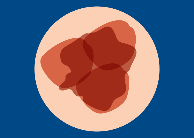
What is Melanoma?
Understanding melanoma can be difficult. Many questions can arise: How do I prevent it? How is it diagnosed? Why is it so deadly? What other information do I need to know to keep me and my family safe? Explore Melanoma Basics and the Types of Melanoma below.


The Basics
Melanoma is a type of cancer, most often of the skin. It occurs in melanocytes – the cells that produce the pigment melanin that colors the skin, hair and eyes. These cells also make moles, or nevi. Having moles can be a risk factor for melanoma, but it is important to remember that most moles DO NOT become melanoma.1
Unlike other cancers, most melanomas can often be seen on the skin, making it easier to detect in its early stages. If left undetected, however, melanoma can spread to distant sites or distant organs. This is referred to as metastatic melanoma. When melanoma spreads, it most commonly spreads to the liver, lungs, bones and brain, making treatment more difficult.
View our You and Melanoma Animated Patient Videos to learn more about melanoma basics, melanoma prevention and melanoma risk factors, or view the Spanish version of Usted y Melanoma Animated Patient Videos.

What Causes Melanoma?
Research suggests that nearly 90% of cutaneous melanoma cases can be linked to exposure to ultraviolet (UV) rays – either from natural sources, like the sun, or from artificial sources, like indoor tanning beds sheet.2 However, since melanoma can occur in all melanocytes throughout the body, even those that are never exposed to the sun, UV light cannot be solely responsible for all diagnoses, especially mucosal and ocular melanoma cases. Current research points to a combination of family history, genetics and environmental factors that are to blame.
Taking steps to prevent melanoma is the best first step in protecting yourself and your skin. It is important to learn about all of the risk factors.

Why Is Melanoma So Serious?
Melanoma is the most serious type of skin cancer because it can spread to lymph nodes and distant organs. In 2023, nearly 187,000 Americans are expected to be diagnosed with melanoma. Of these, more than 97,600 will be diagnosed with invasive (Stage I, II, III or IV) melanoma and over 89,000 will be diagnosed with melanoma in situ (Stage 0).3 Read more melanoma facts and statistics here.
Additionally, Metastatic melanoma is a term used for melanoma that has spread beyond the original site to the lymph nodes or to distant organs.

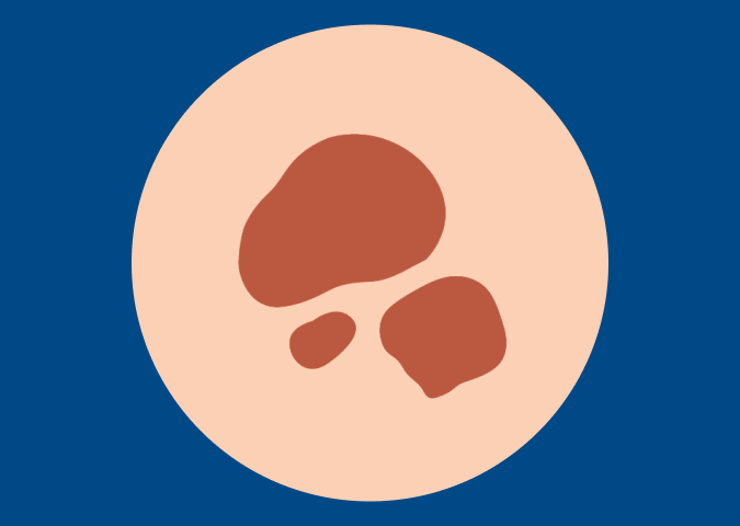
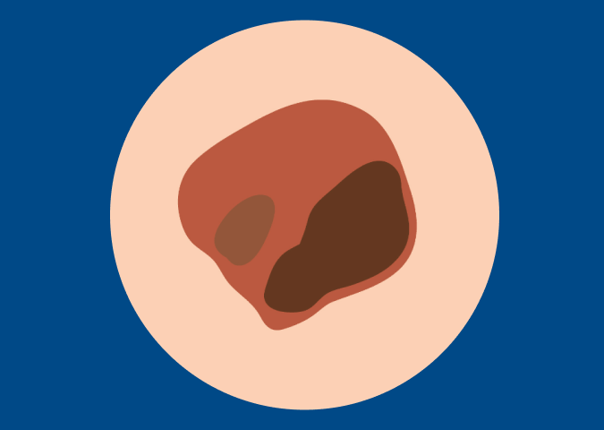
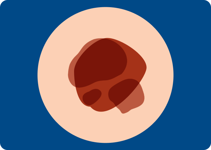
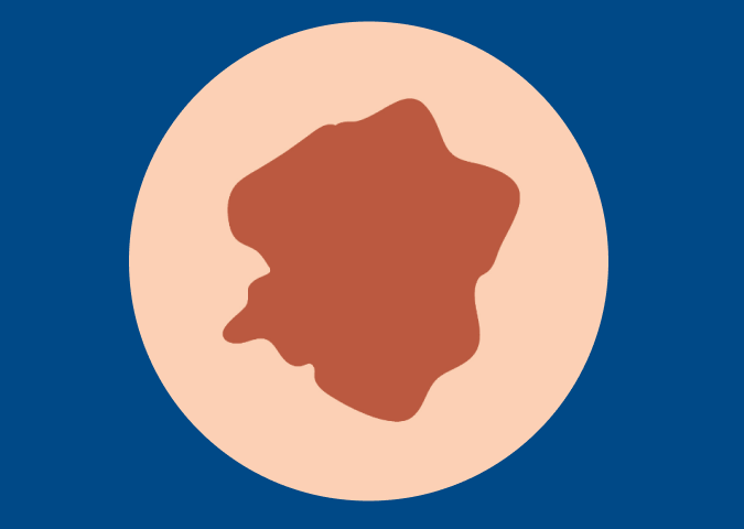
What Does Melanoma Look Like?
Melanoma can look different from person to person, but if you suspect that a spot on your skin fits the following descriptions, talk to your dermatologist right away. You can start here, but note that not all skin cancers and melanomas fall into simple categories.

A Change on the Skin
This could be a new spot, or a change in color, shape or size of a current spot
A New Spot, Sore or Mole
If it doesn’t heal or becomes painful and tender
A Mole That is Irritated
Especially take concern if it starts to itch or bleed
A Firm Red Lump Appears
A firm red lump that bleeds or appears crusty
A Change in in Texture or Look
A spot, sore, mole or lump that looks shiny, waxy, smooth or pale
A Dry, Rough Red Spot
A flat, red spot that is rough, dry or scaly

A Dark Spot or Streak
Found under a fingernail or toenail (that hasn’t come from previous trauma to the nail)³
ANVP and Glossary of Terms
We’ve compiled a list of terms commonly used by oncologists, dermatologists and pathologists that you may see on your patient record or pathology report. Still have questions? Email the MRF nurse or visit the Patient Forum to find answers.

Citations
Content last reviewed: August 20, 2020
North American Association of Central Cancer Registries (NAACCR). Estimated deaths are based on 2003-2017 US mortality data, National Center for Health Statistics, Centers for Disease Control and Prevention.
Lucas RM, McMichael AJ, Armstrong BK, Smith WT. Estimating the global disease burden due to ultraviolet radiation exposure. Int J Epidemiol. 2008;37(3):654-667
National Cancer Institute, Division of Cancer Epidemiology and Genetics, https://moles-melanoma-tool.cancer.gov/