
March 31, 2026
About the MRF’s CURE OM Initiative.
CURE OM (the Community United for Research and Education of Ocular Melanoma) is the MRF’s initiative to increase awareness, education and research funding for ocular melanoma, while improving the lives of people affected by this disease. Founded in 2011, the CURE OM initiative excels because of the hard work and dedication of its Patient and Scientific Steering Committees and the generosity of our community. Show your support with a tax-deductible donation to the CURE OM initiative today!
Understanding Ocular Melanoma.
Many questions can arise: How do I prevent it? How is it diagnosed? Why is it so deadly? What do I need to know to keep me and my family safe? Our team has assembled the sections below to help you navigate your or your loved one’s ocular melanoma journey.


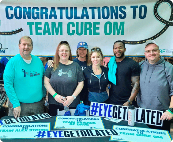
#EYEGETDILATED
OM Patient & Caregiver Resources
Education
The CURE OM initiative is committed to being the leading provider of ocular melanoma education, resources and support. We partner with OM clinicians and researchers to ensure our OM patient and caregiver community always receives the best, most accurate information.
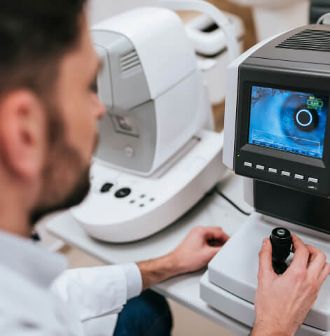
Through the #EyeGetDilated campaign, CURE OM aims to increase awareness by promoting the importance of early detection. OM is most often detected during a routine, dilated eye exam.
Eye on a CURE: Patient & Caregiver Global Symposium – CURE OM hosts an annual in-person educational symposium with major academic medical centers. These meetings provide an opportunity to learn from leaders in ocular melanoma and network with other OM patients and caregivers. Experts provide updates on diagnosis, treatment, clinical trials and research. View past Symposium recordings and learn about upcoming meetings here:

Find Support
CURE OM offers virtual support groups to individuals diagnosed with primary and metastatic ocular melanoma (OM). These virtual sessions include a mix of educational and wellness information as well as space to connect with other patients for emotional support and companionship. We request that this be a safe space away from judgment or any direct medical advice. For more information about participating in a virtual OM support group please email Miriam Kadosh, Director of Patient Engagement and Education at education@melanoma.org.
Find an OM Specialist: Use the Treatment Center Finder to explore ocular melanoma treatment centers. Please note, this is not a doctor recommendation list, simply a list of OM specialists to get you started. The Eye Cancer Network also provides a list of specialists in the US and more than 50 countries around the world.
Form a CURE OM Miles for Melanoma Team
Miles For MelanomaAttend a Gala
GalasStart a CommUNITY Fundraiser
CommUNITY FundraisingVISION Registry Discussions Boards
VISION PlatformAdvocate at State & Federal Level
Advocate for MelanomaOM Research
The CURE OM initiative is leading the ocular melanoma (OM) field forward. We are committed to accelerating the development of effective treatments and, ultimately, a cure for this disease. Our scientific initiative includes research grants, a patient registry and biannual international scientific meetings. These efforts ensure continued collaboration, support and coordination to move OM research forward.
To date, the MRF has funded over 28 ocular melanoma grants nearly over $2.6 million. To learn about upcoming CURE OM research opportunities, visit our Research Grants RFP Page. To learn more about past uveal melanoma research grantees, visit our Funded Research Page and filter by melanoma type → Ocular Melanoma.
Researchers/Clinicians: to learn more about the CURE OM research opportunities, please contact CUREOM@Melanoma.org.
CURE OM hosts regular global science meetings to facilitate interdisciplinary and innovative collaboration focused on finding effective treatments and, ultimately, a cure for ocular melanoma. Each meeting includes a diverse group of speakers and participants from around the world. To learn more about upcoming & past CURE OM scientific meetings, search our Scientific Meetings Page.
Vision Platform
In 2021, CURE OM launched the VISION Registry, a global patient-reported registry to support advances in ocular melanoma research and treatment development. Designed with the guidance of the Registry Steering Committee, the VISION Registry will inform patient-centered research initiatives focused on policy, patient preferences and standards of care. In addition to collecting invaluable data about diagnosis, disease progression, treatment choice and outcomes, geography and environmental factors, the registry supports the collaboration of patients, caregivers, clinicians, researchers, and the pharmaceutical industry to find a cure together.
In 2023, the MRF’s CURE OM initiative expanded the VISION Registry to create the VISION Platform. The goal of the VISION Platform, is to unite the global ocular melanoma (OM) community by providing an interactive and innovative central place where dispersed patients and caregivers can access disease-specific resources and tools and share their knowledge and experiences with each other and to advance research. The VISION Platform continues to grow in collaboration with OM patient and community input and feedback and currently encompasses the following key components:
The CURE OM Clinical Trial Finder will help you understand which emerging ocular melanoma treatments may be an option for you. Clinical trials offer OM patients the opportunity to receive care that may not yet be standard. You must be logged in via the VISION Registry to access the OM Clinical Trial Finder.